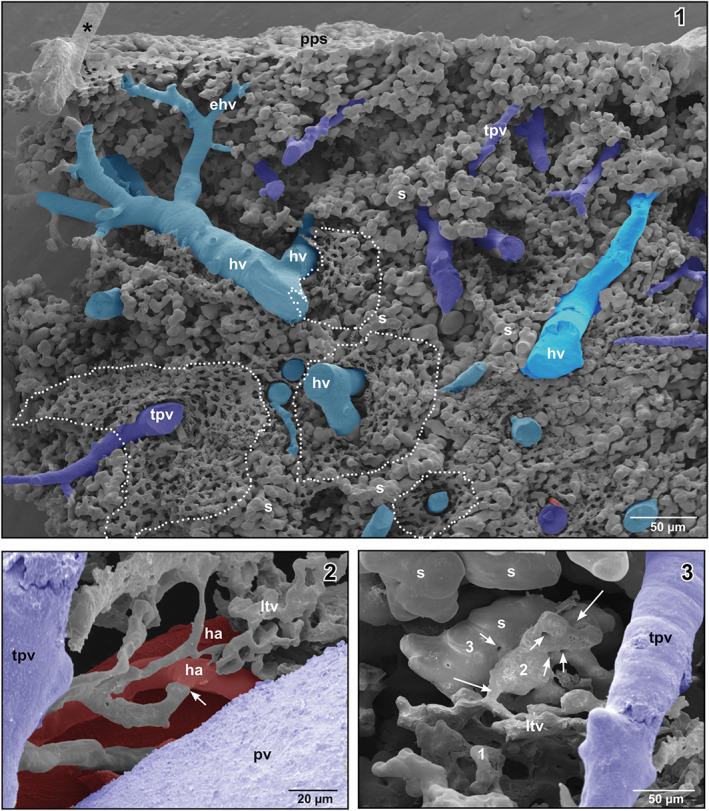
FIGURE 7.

(1) Honeycomb‐like microvascular meshwork around hepatic venules (hv) and terminal portal venule (tpv) (encircled areas). Asterisk marks “conductive bridge.” (2) Arterial supply of the lymphoid tissue vasculature (arrow). (3) Gradual transition (1–3) of the lymphoid tissue vasculature into sinusoids (s) (large arrows). Note the “holes” indicating non‐sprouting angiogenesis (small arrows)
