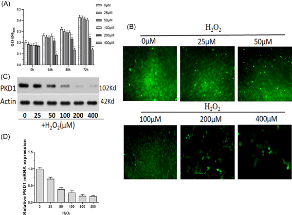
Figure 1.

Reduced expression of PKD1 and cell viability in BM‐MSCs treated with H2O2. Primary rat BM‐MSCs were treated with 0–400 µM H2O2. (A) Cell viability was measured using CCK8. (B) FDA was used to fluorescently label BM‐MSCs. (C) Western blot analysis was performed to measure the protein expression of PKD1. (D) qPCR was performed to measure the mRNA expression of PKD1. Data were expressed as the mean ± SD, n = 3. *p < .05, **p < .01 versus 0 µM H2O2‐treated cells. BM‐MSCs, bone marrow‐mesenchymal stem cells; FDA, Food and Drug Administration; mRNA, messenger RNA; qPCR, quantitative polymerase chain reaction
