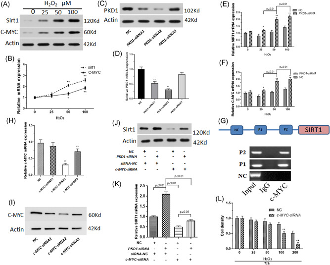
Figure 2.

Sirt1 is targeted by c‐MYC to inhibit PKD1 expression in BM‐MSCs after oxidative stress injury. Primary rat BM‐MSCs treated with 0–100 µM H2O2. (A) The protein expression of Sirt1 and c‐MYC was measured using western blot analysis. (B) The mRNA expression of Sirt1 and c‐MYC was measured by qPCR. n = 3. *p < .05, **p < .01 versus 0 µM H2O2‐treated BM‐MSCs. (C) Western blot analysis was performed to measure PKD1 protein expression in BM‐MSCs transfected with RNA interference fragments (siRNA) of PKD1. (D) PKD1 mRNA expression of BM‐MSCs transfected with RNA interference fragments of PKD1 was detected by qPCR. (E, F) mRNA expression of Sirt1 and c‐MYC in PKD1‐siRNA transfected BM‐MSCs simultaneously treated with 0–100 µM H2O2. (G) ChIP assay was used to explore whether c‐MYC may bind to the promoter of Sirt1 to upregulate its expression. (H, I) Western blot analysis and qPCR assays were used to measure the c‐MYC expression of BM‐MSCs transfected with RNA interference fragments (siRNA) of c‐MYC. (J, K) Western blot analysis and qPCR assay was performed to measure Sirt1 expression in BM‐MSCs transfected with PKD1‐siRNA and/or c‐MYC‐siRNA undergoing oxidative stress induced by 100 µM H2O2. (L) Cell viability of BM‐MSCs transfected with PKD1‐siRNA and/or c‐MYC‐siRNA undergoing oxidative stress was measured using CCK8. Data are expressed as the mean ± SD, n = 3. *p < .05, **p < .01 versus negative control RNA fragment (NC) transfected cells. BM‐MSCs, bone marrow‐mesenchymal stem cells; ChIP, chromatin immunoprecipitation; mRNA, messenger RNA; qPCR, quantitative polymerase chain reaction
