
Figure 4.
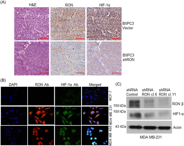
RON knockdown inhibited HIF‐1α expression in the human pancreatic tumor xenograft and RON, HIF‐1α co‐expression in TNBC cells. (A) RON and HIF‐1α expression was analyzed in the BXPC3 control and RON knockdown xenograft FFPE tissue. High RON and HIF‐1α expression was detected in the BXPC3 control xenograft tissue. However, RON knockdown xenograft tissue exhibited a significantly decreased HIF‐1α expression. Scale bar: 100 µm. (B) Immunofluorescence analysis was done on the invasive MDA MB 231, MDA MB 468 TNBC cells, and noninvasive MCF‐7 non‐TNBC cells. RON and HIF‐1α co‐expression was detected in the TNBC cells but not in the non‐TNBC cells. (C) RON and HIF‐1α protein expression was analyzed by Western blot analysis in the MDA MB 231 vector control and RON knockdown clones. RON and HIF‐1α are expressed in the vector control cells, but RON knockdown significantly reduced HIF‐1α expression [Color figure can be viewed at wileyonlinelibrary.com]