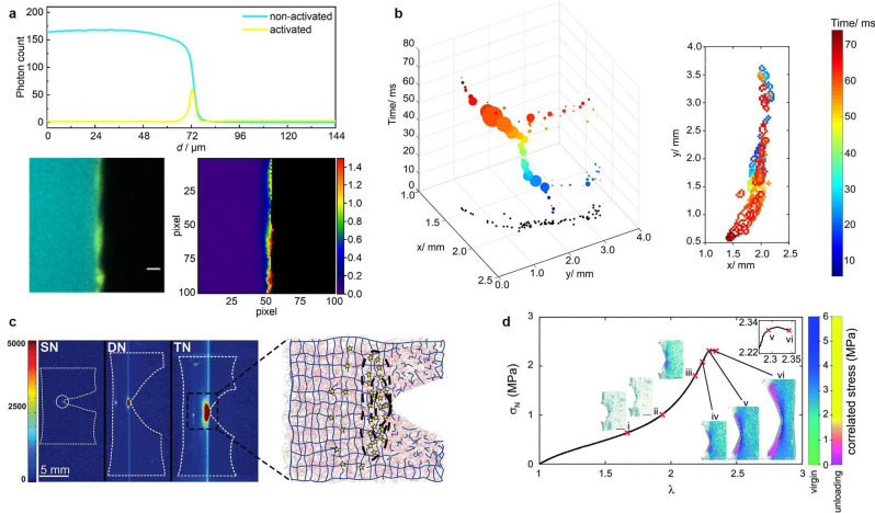
Figure 2.
A selection of bond scission mapping visualizations in OFP‐crosslinked polymer networks: (a) Overlay of CLSM micrographs showing non‐activated (cyan) and activated (yellow) BTD OFP in crosslinked rubber networks, and pixel‐by‐pixel contour map representation of left panel showing the percentage of activated relative to remaining non‐activated BTD upon fracture. Reproduced from Ref. [18]. Copyright 2021, the authors. (b) Progression and extent of a crack front in time and space for dioxetane‐crosslinked PMMA networks under chloroform ingress and swelling‐induced mechanoluminescence. Reproduced from Ref. [19]. Copyright 2017, American Chemical Society. (c) Intensity‐based mapping of bond scission in notched samples of single, double, and triple networks, containing dioxetane crosslinker in the first network, around the tip of a propagating crack and schematic of the bond scission mechanism. Reproduced from Ref. [20]. Copyright 2014, American Association for the Advancement of Science. (d) Stress map around the tip of a propagating crack in acrylate networks crosslinked with spiropyran converted to different merocyanine isomers. Reproduced from Ref. [13]. Copyright 2021, Royal Society of Chemistry.

