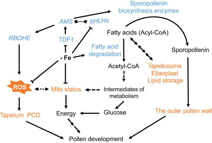
Figure 10.

A model of molecular and cellular responses to Fe deficiency in the tapetum.
In Fe‐deficient anthers, TDF1, AMS, and bHLH089/091/010 are downregulated and lead to many defects in the tapetum and pollen. The downregulation of AMS might alter the expression of RBOHE and genes encoding sporopollenin biosynthesis enzymes. The downregulation of RBOHE reduces ROS production, which may affect tapetum PCD. The change in mitochondrial status possibly results from the decrease in Fe‐dependent mitochondrial protein function. Mitochondrial (Mito) status is crucial for tapetum function in pollen development, as shown in our previous study (Chen et al., 2019). Fe deficiency facilitates fatty acid degradation and fatty acid metabolic flux with reduced lipid storage, and the produced acetyl‐CoA can be used as an intermediate for carbon metabolism and as an energy source to compensate for mitochondrial dysfunction. Taken together, Fe deficiency in anthers triggers extensive transcriptome reprogramming, which might contribute to several tapetum defects, and Fe insufficiency might have impacts on Fe‐dependent proteins and mitochondrial status. These changes in molecular and cellular processes result in disrupted pollen development. The blue font represents genes/proteins involved in molecular regulation. The orange font indicates cellular processes that are defective under Fe deficiency. The line ending in circles represents protein interactions.
