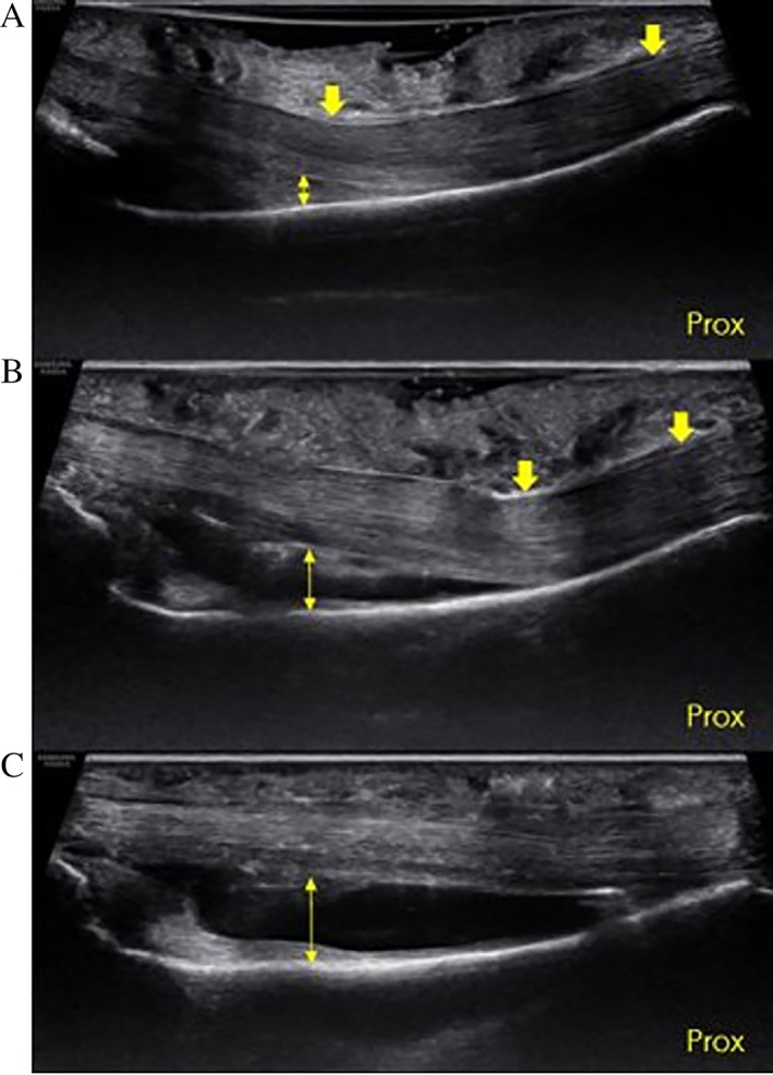
Figure 6.

Ultrasound image of a cadaveric specimen. Long axis view of long finger showing the proximal–distal extent of the proximal phalanx (Prox, proximal). A, Intact A2 pulley (down arrows) in the crimp grip position using a gel standoff. Note how the superficial part of the tendons deflect at the distal end of A2, and the TP distance (double‐headed arrow) is greater than 0 at distal A2 under normal conditions. B, Same specimen following US guided release of the distal 50% of A2 (down arrows). The point of tendon deflection has moved proximally, and the TP distance (double‐headed arrow) has increased. C, Same specimen following US guided release 100% of A2. The bowstringing has increased and the tendon is completely separated from the palmar aspect of the phalanx (double‐headed arrow).
