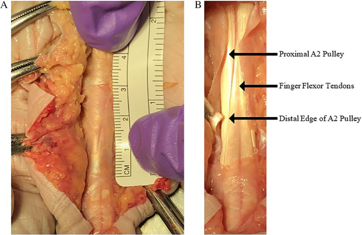
Figure 7.

Cadaveric image of the A2 pulley. A, The proximal fibers of A2 blend with the A1 pulley, creating a challenge in directly distinguishing the transition zone between the two pulleys. B, The arrows point to the proximal A2 pulley, flexor tendon, and the distal edge of the A2 pulley. Note that the leading edge is thickened in comparison to the proximal portion. The thinning of the proximal portion of the A2 pulley creates difficulty in identifying it with ultrasound imaging, a situation that can be improved with a high‐frequency transducer.
