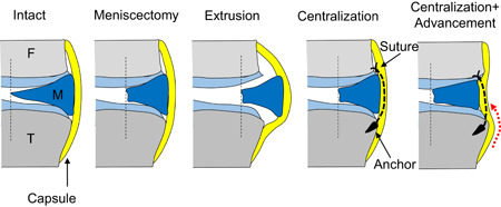
Figure 1.

Experimental settings. Intact—intact lateral meniscus (F: femur, M: meniscus, T: tibia); Meniscectomy—inner half of the posterior half meniscus was removed; Extrusion—posterior meniscus was dislocated laterally by transecting the posterior root and the meniscotibial ligament; Centralization—centralization procedure using one or two anchors; Centralization + advancement—centralization with capsular advancement (red dotted line) using two anchors, which moved the inner margin of the meniscus to the original position of the intact meniscus [Color figure can be viewed at wileyonlinelibrary.com]
