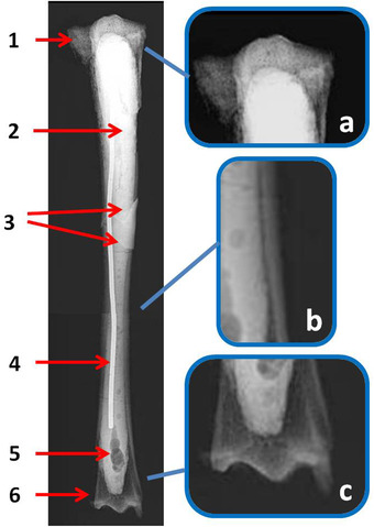
Figure 4.

Candiacervus major, Bate cave (Rethymnon), tibia (specimen number 28 in Capasso Barbato & Petronio 1986). Radiological analysis in craniocaudal view. Wide bone reabsorption area in the proximal (1) and distal (6) epiphyses. The entire medullary cavity has been filled with a sealing material (2) showing air bubbles (5) and incorporating a metal pin (4) in order to strengthen the bone. This suffered a post mortem fracture whose breaking lines can be seen (3). On the right, some enlarged details of the epiphyses (a, c) and diaphysis wall (b) show the rarefied status of the bone tissue.
