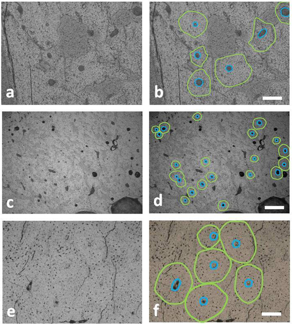
Figure 6.

Secondary osteons in the diaphyseal bone of the tibia (a,b) and metatarsal bone (c,d) of Candiacervus major and metatarsal bone of Candiacervus ropalophorus (e,f). The green line identifies the bone lamella delimiting the osteon area and the blue line indicates the perimeter of the Haversian canal. Bar = 80 μm. (transmitted light microscope; (a) PSC 1848; (c) PSC1867; (e) PSC1873).
