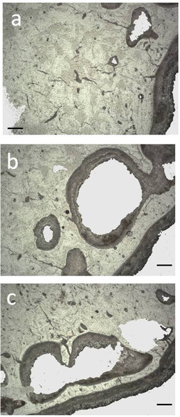
Figure 7.

Histological pictures from the diaphysis of the Candiacervus major from Bate cave: tibia (a,b), metatarsus (c). Wide multiple cavities inside of bone wall can be noted in the subperiosteal zone (a,c) and subendosteal zone (b). Bar = 80 μm. (transmitted light microscope; (a) PSC1854; (b) PSC1857; (c) PSC1865).
