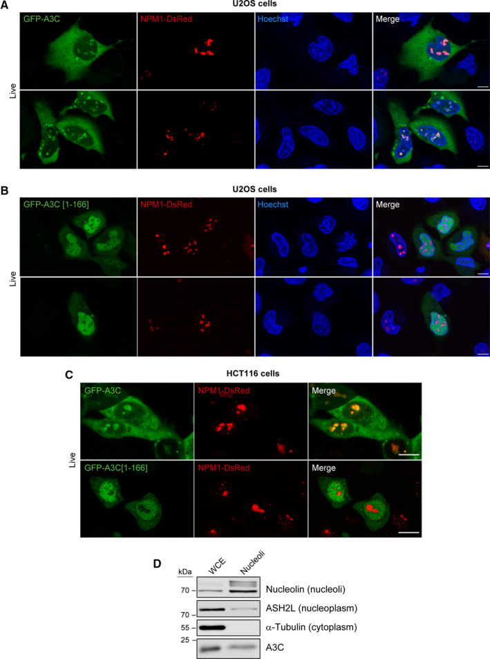
Fig. 10.

The C‐terminal end of A3C is required for its nucleolar location. (A, B) U2OS cells were co‐transfected with hNPM1‐DsRed.cmv and either GFP‐hAPOBEC3C‐V5 (panel A) or GFP‐hAPOBEC3C[1‐166]‐V5 (panel B). The cells were analyzed 24 h post‐transfection using a confocal microscope. Nuclei were stained with Hoechst 33342. (C) As panels A and B but using HCT116 cells instead of U2OS cells. (D) Western blot indicating the presence of A3C in whole cell extracts (WCE) and purified nucleoli from HCT116 cells. Scale bars in panels A to C, 10 µm.
