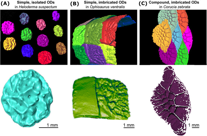
Fig. 3.

Shapes and distribution pattern of some dorsal skin osteoderms (ODs) in three species of lizards. Dorsal views of the distribution of neighbouring osteoderms (top) and the morphology of isolated osteoderms (bottom) in (A) Heloderma suspectum (Helodermatidae), (B) Ophisaurus ventralis (Anguidae), and (C) Corucia zebrata (Scincidae).
