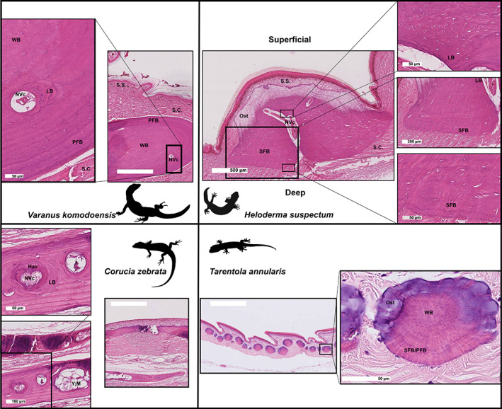
Fig. 4.

Histological overview of osteoderms from Varanus komodoensis, Heloderma suspectum, Tarentola annularis, and Corucia zebrata, demonstrating diversity of osteoderm size, tissue characteristics and bone fibre patterning. All images stained with haematoxylin and eosin. Hav, Haversian structure; LB, lamellar bone; NVc, neurovascular canal; Ost, osteodermine; PFB, parallel‐fibred bone; S.C., stratum compactum; SFB, Sharpey‐fibre bone; S.S., stratum superficiale; WB, woven bone; Y; M, yellow marrow. Scale bars: main images all 200 μm; higher magnifications as indicated. Varanus komodoensis and Heloderma suspectum from the same specimens as Kirby et al. (2020).
