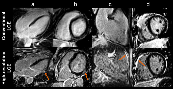
FIGURE 3.

Late gadolinium enhancement (LGE) images from conventional LGE (top row) and high‐resolution LGE (bottom row) in patients with myocardial infarction defined by sub‐endocardial LGE (a), myocarditis with sub‐epicardial LGE (b), arrhythmogenic right ventricular cardiomyopathy (c), and repaired tetralogy of Fallot with LGE at the surgical scar (d) (for all, LGE is designated by the yellow arrows). These images are adapted from references 11, 12, 16 with the courtesy of Prof. Cochet.
