
FIGURE 8.
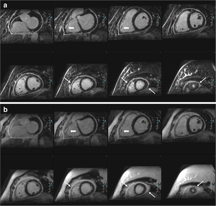
Comparison of (a) HR‐LGE with fat‐water separation (DIXON) and (b) conventional 2D LGE in a female patient with pericarditis. DIXON‐based fat suppression enables excellent delimitation of the enhanced pericardium against the epicardial fat. In direct comparison, the pericardium can hardly be identified in several areas in the HR‐LGE views and worse in the conventional LGE views (thin arrows: eg, along the right ventricle, close to the apex). Moreover, 3D water LGE imaging allows for excellent depiction of small details such as the trabeculae of the right ventricle (bold arrows). Reprinted from reference 108 with permission from Springer.