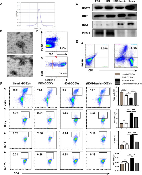
FIGURE 2.

Mouse dendritic cell (DC)‐released extracellular vesicles (DCEVs) identification and effect on Th cell subsets differentiation. EVs were obtained from cell culture supernatants and identified by scanning electron microscopy (SEM), Western blot, flow cytometry (FCM), and nanoparticle microscopy tracking analysis (NTA), respectively. A: NTA; B: SEA; C: expression of heme oxygenase (HO)‐1, MHC II, heat shock protein (HSP)70, and CD81 were examined by Western blot; and D: expression of phosphatidylserine (PS; Annexin V+) in EVs was detected by FCM. Mouse CD4+ naïve T cells were cocultured in vitro with enhanced green fluorescent protein (EGFP)+DC‐derived EVs for 2 d. E: Proportion of EGPF+T cells was detected by FCM. Mouse CD4+ naïve T cells were cocultured in vitro with different stimulus‐induced DCEVs for 6 d. F: Proportion of regulatory T cells (Treg; CD4+ CD25+ forkhead box P [Foxp]3+), Th1 (CD4+ IFN‐ γ+), Th2 (CD4+ IL‐13+), and Th17 cells (CD4+ IL‐17A+) were detected by FCM. For statistical analysis of FCM, data are pooled from three independent experiments. The data are shown as mean ± sd, *P < 0.05; **P < 0.01; ***P < 0.001; ns, not significant
