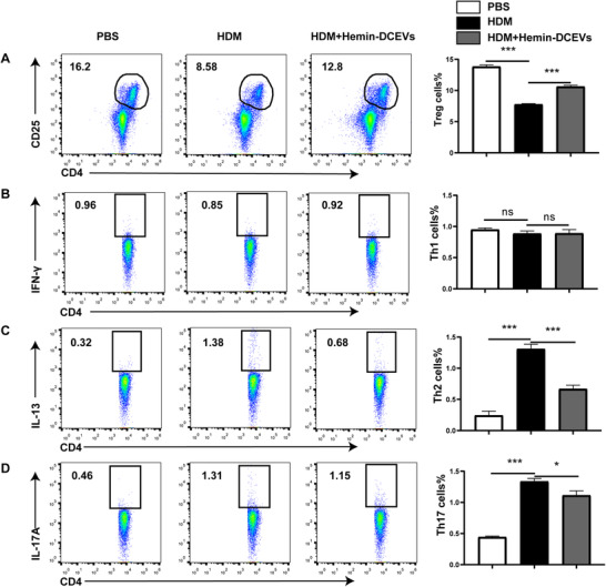
FIGURE 5.

Hemin‐treated dendritic cell (DC)‐released extracellular vesicles (DCEVs) affect proportion of Th cells in mouse mediastinal lymph node (MLNs). House dust mite (HDM)‐sensitized and challenged mice were instilled intranasally with 100 μg hemin‐DCEVs at 1 d before sensitization and challenge (HDM + hemin‐DCEVs group). A: flow cytometry (FCM) of regulatory T cells (Tregs; CD4+ CD25+ forkhead box P [Foxp]3+) in MLNs; B: FCM of Th1 (CD4+ IFN‐ γ+) in MLNs; C: FCM of Th2 (CD4+ IL‐13+) in MLNs; and D: FCM of Th17 (CD4+ IL‐17A+) in MLNs. For statistical analysis of FCM, data are pooled from three independent experiments with three mice each group. The data are shown as mean ± sd, *P < 0.05; ***P < 0.001; ns, not significant
