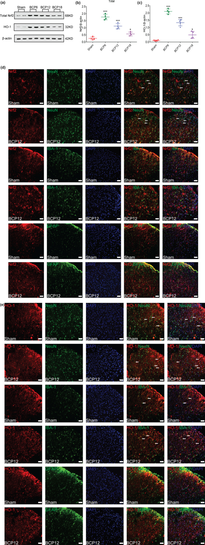
FIGURE 3.

Dynamic changes in nuclear factor erythroid 2 (NFE2)‐related factor 2 (Nrf2) and heme oxygenase‐1 (HO‐1) expression and cellular localization after tumour inoculation. (a–c) Western blot analysis showed that after tumour inoculation, Nrf2(a and b) and HO‐1(a and c) expression levels in the spinal cord of the model group were significantly increased compared with those in the sham group on days 6, 12 and 18 after the operation (*p < 0.05,***p < 0.001 vs. sham group; n = 4, one‐way ANOVA). (d and e) Immunofluorescence results showed that in the dorsal horn of the spinal cord of bone cancer pain (BCP) rats, Nrf2 (red) was primarily expressed in neurons (green) rather than astrocytes (green) or microglia (green), whereas HO‐1 (red) was primarily expressed in neurons (green) and partially in microglial cells (green), lacking colocalization with astrocytes (green). Lumbar enlargements were collected on day 12 after the operation or tumour inoculation. Sections were counterstained with DAPI (blue) to label cell nuclei. The white arrows indicate colocalization of Nrf2 and HO‐1 with NeuN (neuronal nuclei, neuronal‐specific marker), GFAP (glial fibrillary acidic protein, astrocyte specific marker) and Iba‐1 (ionized calcium binding adapter molecule 1, microglial‐specific marker)‐immunoreactive cells in the spinal dorsal horn, respectively; n = 4. Scale bar = 50 μm. n represents the number of experimental animals in each group
