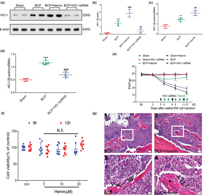
FIGURE 6.

Spinal heme oxygenase‐1 (HO‐1) activation is essential for the regulation of bone cancer pain (BCP). (a–d) After treatment with HO‐1 agonist or inhibitor, HO‐1 expression in the spinal cord was observed. Rats were killed after 12 days of BCP to collect spinal cord tissue. Hemin and HO‐1 siRNA further stimulated and inhibited the expression of HO‐1 in the spinal cord of rats with BCP, respectively (***p < .001 vs. sham group; ## p < .01, ### p < .001 vs. BCP group; n = 4, **p < .01,***p < .001 vs. sham group; ## p < .01, ### p < .001 vs. BCP group; n = 5, one‐way ANOVA). (e) The effects of HO‐1 agonist (hemin) and HO‐1 inhibitor (HO‐1 siRNA) on the paw withdrawal threshold (PWT) in rats with BCP were observed within the indicated duration after inoculation(*p < .05, ***p < .001 vs. BCP group; n = 6, two‐way repeated‐measures ANOVA). (f) Hemin was treated with the indicated concentration for 6 hr and 12 hr. A CCK‐8 assay was used to detect Walker 256 cell viability. N.S stands for not statistically significant (p > .05 vs. control group, two‐way repeated‐measures ANOVA). (g) Representative images of haematoxylin‐eosin staining showing cancer cell infiltration (cells within the dotted lines) and bone resorption pits (black arrows) on the trabecular surface in the tibial marrow cavity of the BCP (g (1, 2)) and BCP +hemin (g (3, 4)) groups; n = 10. Scale bars: 200 μm (top row), 50 μm (bottom row). Rats were killed on BCP 12 days to collect tibia tissues. n represents the number of experimental animals in each group
