
Figure 5.
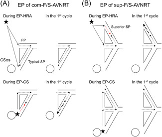
Schematic illustration of the putative mechanism of shortening of retrograde conduction over the slow pathway (SP) after atrial EP of com‐F/S‐AVNRT (A) and sup‐F/S‐AVNRT (B). The thin arrows show the directions of propagation in the atrium, and the black and red dotted arrows show the orthodromic and antidromic directions of propagation in the AV nodal pathways. The bold, dotted arrows illustrate the accelerated retrograde conduction over the atrial side of the typical or superior SP. (A) When antidromic penetration into the typical SP is deeper during EP from the proximal coronary sinus (EP‐CS) than from the high right atrium (EP‐HRA; left panel), the retrograde conduction time over the typical SP in the first cycle is shorter after EP‐CS than after EP‐HRA (right panel), causing an unequal response. (B) When antidromic penetration into the superior SP is deeper during EP‐HRA than during EP‐CS (left panel), the retrograde conduction time over the superior SP in the first cycle is shorter after EP‐CS than after EP‐HRA (right panel), causing an unequal response. See text for details. CSos, ostium of coronary sinus, FP, fast pathway. The asterisks indicate the EP site in the HRA or proximal CS. AVNRT, atrioventricular nodal reentrant tachycardia; CS, coronary sinus; EP, entrainment pacing; HRA, high right atrium