
Fig 1.
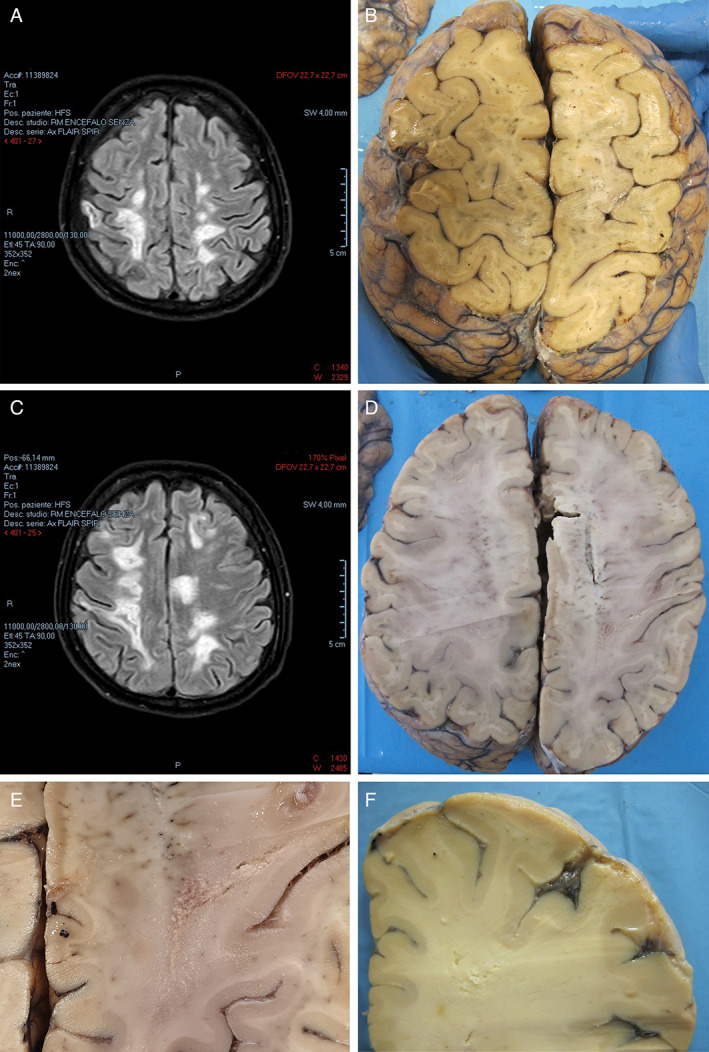
MRI (A, C) and gross (B, D‐F) findings of the cerebral hemispheres. (A, C) Axial FLAIR images taken on day 88 after first hospital admission show hyperintense areas in the cerebral white matter. (B, D) Horizontal brain slices corresponding to the imaging views (A, C) show the appearance of white matter lesions. (E, F) Details of lesions at higher magnifications are shown. Panels E and F correspond to selected area of slice D and taken at another slice.