
Fig 3.
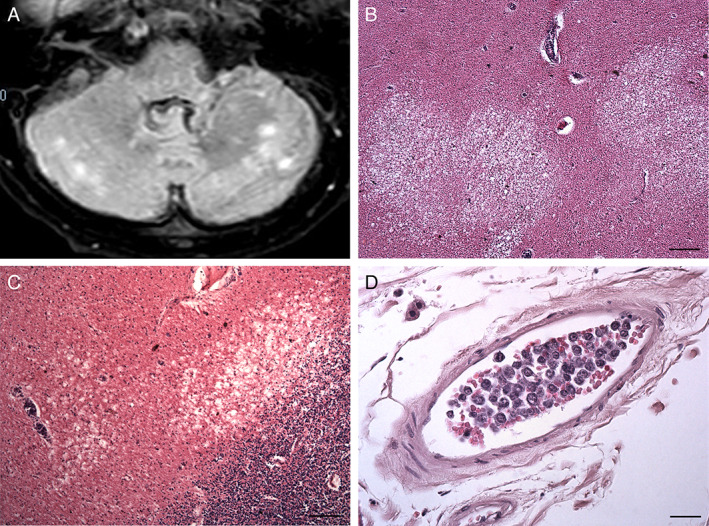
MRI (A) and histological (B‐D) findings of the cerebellum. (A) An axial FLAIR Spir image taken on day 88 after admission shows hyperintense lesions in the cerebellar white matter. (B, C) Necrotic lesions are observed in the cerebellar white matter. (D) A leptomeningeal medium‐sized vein is filled with large round cells. Scale bars, 200 μ (B), 100 μm (C), 25 μm (D).