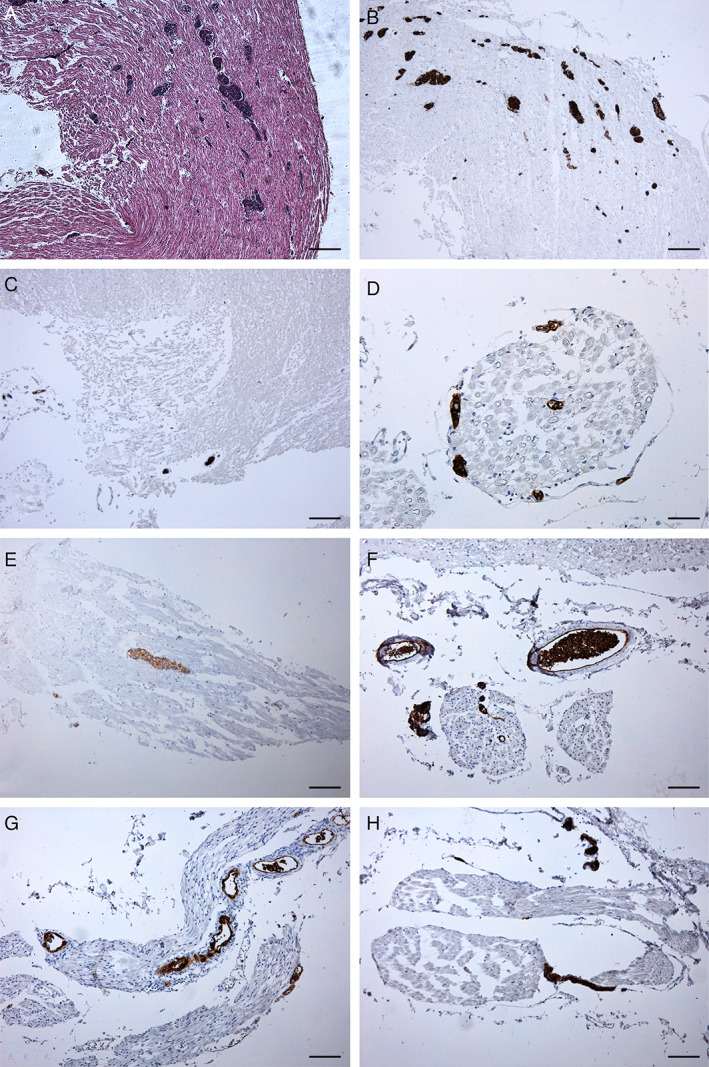
Fig 6.

Findings of the cranial nerves on HE staining (A) and CD20 immunohistochemistry (B‐H). (A) Small vessels occluded by round cells are observed in the right oculomotor nerve trunk. (B‐H) Vessels occluded by CD20‐positive malignant B cells are visible in the right oculomotor (B), left (C, D) and right (E) trigeminal, left glossopharyngeal (F), right vagal (G), and right hypoglossal (H) nerves. Scale bars, 200 μm (A‐C), 50 μm (D), 100 μm (E‐H).
