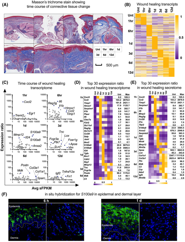
FIGURE 3.

Extraction of transcripts related to wound healing using a gene ontology term. A, Photomicrographs of Masson's trichrome staining of incised hind paw tissues. B, Heatmap of the transcripts related to wound healing. C, The differentially expressed genes in the transcripts related to wound healing at each timepoint with several representative genes labeled. For the scatter plot, the expression ratio between each timepoint and untreated control is plotted versus average sFPKM using logarithmic scales. D, The top 30 differentially expressed transcripts related to wound healing. E, The top 30 differentially expressed transcripts of the wound‐healing secretome. Note the stepwise temporal patterns of transcript increases in B, D and E. F, Photomicrographs of in situ hybridization for S100a9 (green) and DAPI (blue) in the incised hind paw at 6 hours and 1 day after incising. Left panels in each timepoint shows the epidermis and the upper layer of dermis and right panels in each timepoint shows the middle layer of dermis. The vast majority of S100a9 in situ signal is located in the epidermal layers. d, day(s); hr or h, hour(s); Unt, untreated control
