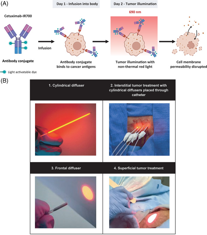
FIGURE 1.

Mechanism of action and RM‐1929 photoimmunotherapy overview. (A) A tumor‐targeted antibody is conjugated with a light‐activatable dye. Following infusion into the body, the tumor is illuminated with non‐thermal red light (690 nm), leading to anticancer activity mediated by biophysical processes that damage the membrane integrity of cells. (B) Tumor illumination is performed 24 ± 4 h after antibody conjugate infusion. Cylindrical diffusers placed in needle catheters are used to treat interstitial tumors (B1, B2), while frontal diffusers are used to treat superficial tumors (B3, B4). Light illumination of the tumor is administered 24 ± 4 h after RM‐1929 infusion to allow for drug distribution within the tumor. The non‐thermal red light is applied to the tumor using a (1) frontal diffuser for superficial light illumination for tumors ≤1 cm from the skin or mucosal surface or a (2) cylindrical diffuser for interstitial illumination for tumors >1 cm from the skin or mucosal surface. The illumination time for frontal and cylindrical diffusers is 5 min for each treated area. For interstitial illumination, cylindrical diffusers are placed uniformly into the tumor 1.8 ± 0.2 cm apart using 17‐gauge closed‐tipped needle catheters under radiographic or ultrasound imaging
