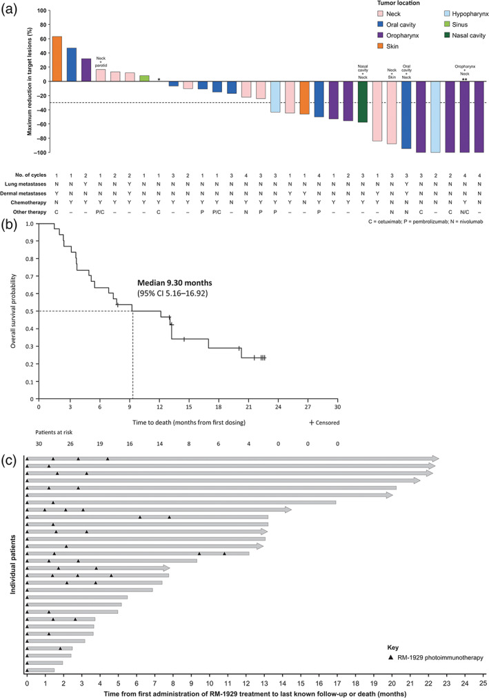
FIGURE 2.

Efficacy outcomes following RM‐1929 photoimmunotherapy in study Part 2 patients (n = 30) with locoregional rHNSCC. (A) Waterfall plot of best change in sum of diameters of target lesions from baseline by central radiology assessment; (B) Kaplan–Meier plot of overall survival; and (C) Swimmer plot of individual patient survival. Arrowed bars represent patients who were censored at last contact date. *One enrolled patient did not have post‐baseline scans. **Lesions completely resolved, and lymph nodes were <10 mm. Target lesions were defined as lesions identified by the investigator that were light illuminated. Non‐target lesions, including lung metastases, did not undergo light illumination and further discussion on change in tumor size of non‐target lesions can be found in Appendix S1. Tumor locations were determined by a sponsor clinical expert based on target and non‐target lesions reported in the study
