Figure 8.

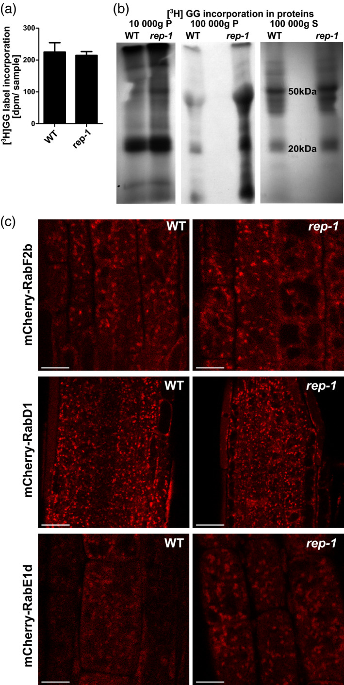
Rab protein prenylation and localization in the rep‐1 mutant.
(a) Quantification of in vivo metabolic incorporation of [3H]geranylgeraniol in plant proteins with a molecular mass of 17–30 kDa. Total lysates were prepared without fractionation and resolved by SDS‐PAGE. Gel regions corresponding to the size of Rab proteins (17–30 kDa) were cut out and solubilized, and their radioactivity was quantified in a scintillation counter. Bars represent the mean of at least three independent biological experiments ± SD.
(b) In vivo metabolic incorporation of [3H]geranylgeraniol in plant proteins. Total lysates were prepared from seedlings of WT and rep‐1/rep‐1 lines cultured on medium containing [3H]geranylgeraniol. Lysates were separated into three fractions, 10 000 g pellet, 100 000 g pellet, and 100 000 g supernatant, resolved by SDS‐PAGE, and analyzed by autoradiography. The results of a representative experiment are shown. Fractions from the 100 000 g pellet showed much higher [3H]GG incorporation, and lower exposure of the same gel is shown. In all lanes except the rep‐1 100 000 g pellet a similar amount of total protein per lane was loaded; in the rep‐1 100 000 g pellet lane a higher amount of protein was loaded.
(c) Localization of selected Rab proteins in the rep‐1 background. mCherry fusions of Rab‐F2b (upper panel), Rab‐D1 (middle panel), and Rab‐E1d (lower panel) were introduced into the rep‐1/rep‐1 line by crossing. Localization in root epidermal cells (upper and lower panels) and root meristematic cells (middle panel) is shown. CSLM images, scale bar = 10 µm for upper and lower panels and scale bar = 20 µm for the middle panel.
