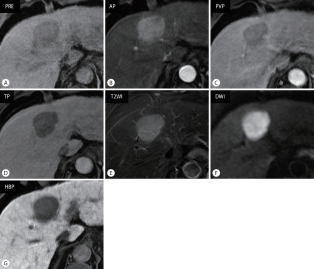
Figure 1.
Typical magnetic resonance imaging findings of hepatocellular carcinoma using hepatobiliary contrast agent in a 61-year-old male patient with chronic hepatitis B. Approximately 4-cm sized mass lesion in liver segment 4, which shows (A) hypointensity in precontrast T1-weighted image, (B) arterial phase hyperenhancement, and (C) portal venous phase (PVP) and (D) transitional phase (TP) washout. It shows (E) moderate hyperintensity in fat-suppressed T2-weighted image and (F) diffusion restriction. (G) In the hepatobiliary phase (HBP) image acquired 20 minutes after contrast administration, the mass is clearly visualized with hypointense lesion in contrast to hyperintense hepatic parenchyma. PRE, precontrast; AP, arterial phase; T2WI, T2-weighted image; DWI, diffusion-weighted image.

