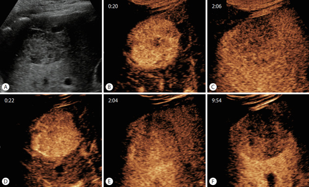
Figure 2.
Typical contrast-enhanced ultrasound findings of hepatocellular carcinoma using SonoVue and Sonazoid. (A) Approximately 4.5-cm sized hypoechoic mass in liver segment 6 on B-mode ultrasound. After administration of SonoVue, the mass shows (B) hyperenhancement in the arterial phase (20 seconds) and (C) mild washout in the delayed phase, but not devoid of enhancement (126 seconds). The same lesion enhanced with Sonazoid also shows (D) arterial phase hyperenhancement (22 seconds) and (E) mild delayed washout (124 seconds), as well as (F) clear hypointensity in the Kupffer phase (approximately 10 minutes after contrast administration).

