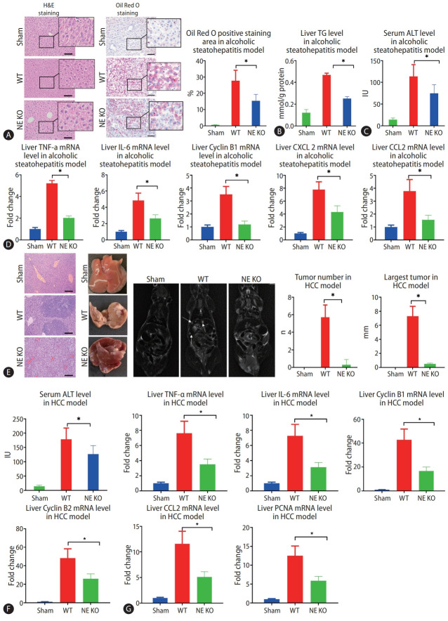
Figure 2.
Lack of neutrophil extracellular traps (NETs) leads to alleviation of alcoholic hepatosteatosis and subsequent hepatocellular carcinoma (HCC) in mice. (A) Mouse liver tissues were visualized by hematoxylin and eosin (H&E) staining and Oil Red O staining after 5 weeks of alcohol treatment (scale bar, 100 μm). (B, C) Liver triglyceride (TG) and serum alanine aminotransferase (ALT) levels from alcoholic hepatosteatosis model mice were determined. (D) Wild type (WT) and neutrophil elastase (NE) knockout (KO) mice underwent alcohol treatment for 5 weeks, and liver levels of tumor necrosis factor (TNF)-α, interleukin (IL)-6, cyclin B1, C-X-C motif ligand 2 (CXCL-2), and chemokine ligand 2 (CCL2) genes were determined by qPCR. (E) Liver tumors from HCC model mice were analyzed by H&E staining (scale bar, 200 μm), photography, and magnetic resonance imaging (MRI) scanning. The arrows indicate the location of tumors in MRI scanning. (F) Serum ALT levels were determined in mouse model of HCC. (G) WT and NE KO mice underwent the procedure to establish the HCC model for 13 weeks, whereupon liver levels of TNF-α, IL-6, cyclin B1, Cyclin B2, CCL2, and proliferating cell nuclear antigen (PCNA) genes were determined by qPCR. qPCR, quantitative polymerase chain reaction. *P<0.05 compared between groups (5–8 mice per experimental group).

