Fig. 1.
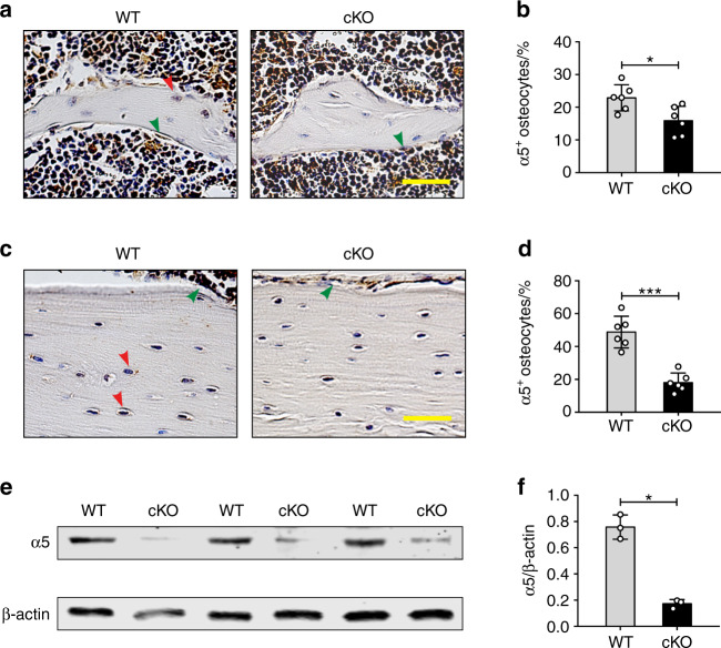
Deletion of integrin α5 gene in osteocytes, but not in osteoblasts in cKO mice. Representative images of integrin α5 immunohistostaining in tibial metaphyseal trabecular bone (a) and midshaft cortical bone (c) of WT and cKO mice. The red arrow shows α5-positive osteocytes and the green arrow shows α5-positive osteoblasts. (a, c) Scale bar: 40 μm. Quantification of integrin α5-positive osteocytes in tibial trabecular bone (b) and cortical bone (d) of WT and cKO mice. n = 6 per group. e, f Integrin α5 deletion was confirmed by western blot of marrow-flushed tibia bone extract samples. n = 3 per group. Mean ± SD. *P < 0.05; ***P < 0.001. Student unpaired t test

