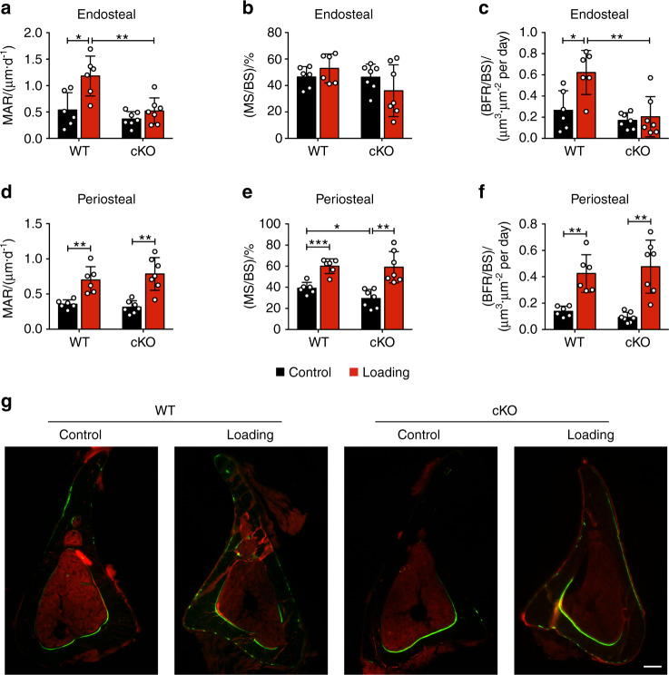
Fig. 4.
Integrin α5 deletion in osteocytes inhibited increased midshaft endosteal osteogenesis by mechanical load. Bone dynamic histomorphometry was conducted on the tibias within diaphyseal 37% cortical VOI, for both loaded and contralateral, unloaded tibias of WT and cKO mice. Mineral apposition rate (MAR) (a, d), mineralizing surface/bone surface (MS/BS) (b, e), and bone formation rate/bone surface (BFR/BS) (c, f) were assessed along endosteal (a–c) and periosteal (d–f) surfaces of all tibias. n = 6 per group. G Representative images of calcein (green) and alizarin (red) double labeling at the 37% VOI for all groups. Scale bar: 200 μm. n = 6 per group. Mean ± SD. *P < 0.05; **P < 0.01; ***P < 0.001. Paired t test was performed for loaded and contralateral tibias and unpaired t test was performed for loaded or control tibias between WT and cKO mice

