Fig. 7.
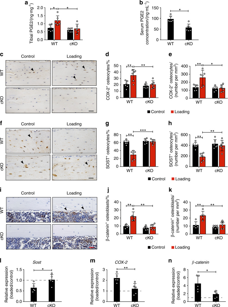
Loading-induced increased PGE2 secretion with a decrease in SOST expression was inhibited in cKO mice. a, b ELISA analysis of PGE2 level in bone marrow-flushed tibial diaphysis (a) and serum (b) after 5-day mechanical loading. n = 6 per group. c Representative COX-2 immunohistostaining (black arrows) and d, e quantification of COX-2-positive osteocytes in diaphyseal 37% cortical bone. Scale bar, 30 μm. n = 6 per group. f Representative SOST immunohistostaining (black arrows) and g, h quantification of SOST-positive osteocytes in diaphyseal 37% cortical bone. Scale bar, 30 μm. n = 6 per group. i Representative β-catenin immunohistostaining (black arrows) and j, k quantification of β-catenin-positive osteoblasts on endosteal surface of diaphyseal 37% cortical bone. Scale bar, 30 μm. n = 5–6 per group. l–n Relative gene expression of Sost (l), COX-2 (m), and β-catenin (n) was determined by RT-qPCR in the tibial diaphysis of WT and cKO mice. n = 5 per group. Mean ± SD. *P < 0.05; **P < 0.01; ***P < 0.001. Paired t test was done for loaded and contralateral tibias and unpaired t test was done for loaded or control tibias between WT and cKO mice

