
Figure 2.
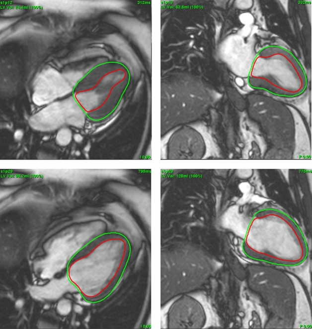
Cardioavascular magnetic resonance LAX images with automated contouring at end systole (top) and end diastole (bottom); 4CV (left) and 2CV (right). 2CV, 2 chamber view; 4CV, 4 chamber view; LAX, long axis.
Official websites use .gov
A
.gov website belongs to an official
government organization in the United States.
Secure .gov websites use HTTPS
A lock (
) or https:// means you've safely
connected to the .gov website. Share sensitive
information only on official, secure websites.
Cardioavascular magnetic resonance LAX images with automated contouring at end systole (top) and end diastole (bottom); 4CV (left) and 2CV (right). 2CV, 2 chamber view; 4CV, 4 chamber view; LAX, long axis.