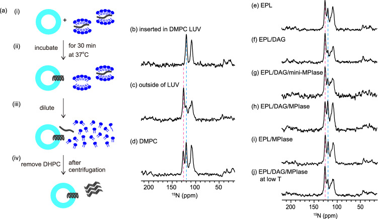
Figure 3.
(a) Procedure for inserting Pf3_24_3 (Fig. 1d) into membranes. (i) Pf3_24_3 (dark gray) solubilized in a DHPC solution was mixed with LUVs composed of various types of lipids. (ii) The mixture was incubated for 30 min at 37 °C. (iii) Then, it was diluted with a buffer solution until the concentration of DHPC reached below the critical micelle concentration of DHPC (1.6 mM). (iv) The supernatant containing DHPC was removed after centrifugation and this step was repeated two times to remove DHPC. We confirmed that Pf3_24_3 outside the membrane was completely precipitated at this stage and not present in the supernatant (Fig. S5). The precipitates were subjected to 15N CPMAS NMR. (b–j) 15N CPMAS NMR spectra of Pf3_24_3. (b) Pf3_24_3 reconstituted into DMPC LUV. The membrane insertion procedure shown in (a) was omitted for this sample. (c) Pf3_24_3 in the aggregates. The sample was prepared as shown in (a) but without using LUVs. Therefore, all proteins were recovered as aggregates. (d–j) The sample was prepared as shown in (a). The LUVs were composed of DMPC (d), EPL (e), EPL/5 mol% DAG (f), EPL/5 mol% DAG/5 mol% mini-MPIase-3 (g), and EPL/5 mol% DAG/1 mol% MPIase (h), EPL/1 mol% MPIase (i). (j) The lipid composition was similar to (h), but all membrane insertion processes were performed below 4 °C. The pink and light blue dashed lines show the chemical shift at 125.6 and 119.6 ppm, respectively.

