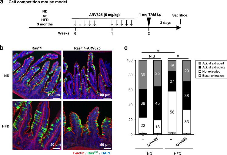
Fig. 5. ARV825 improves apical extrusion of the RasV12-transformed cells in the small intestine of the HFD-fed cell competition model in vivo.
a Timeline of the experimental procedure. Cell competition model mice, LSL-RasV12-IRES-eGFP and villin-Cre-ERT mice, in whom RasV12 expression is induced by tamoxifen treatment (TAM) in a Cre-dependent fashion and traced using simultaneous expression of eGFP18, 27, were fed HFD for 3 months. The black arrow indicates an intraperitoneal treatment with 5 mg/kg ARV825. b, c Immunofluorescence images (b) and quantification analysis (c) of RasV12 cells in the epithelium of the small intestine. Phalloidin (F-actin, red) and RasV12 signals (green) were detected in the small intestine, and DNA was stained using DAPI (blue) (N-RasV12, n = 3 (ND); N-RasV12 + ARV825, n = 3 (ND); N-RasV12, n = 3 (HFD); N-RasV12 + ARV825, n = 3 (HFD)). Scale bars, 50 μm (HFD), 100 μm (ND). *P < 0.05, or not significant (N.S.) by the unpaired two-sided t test.

