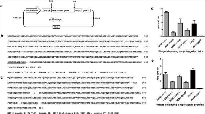
Figure 1.
Display of HER-1/HER-2 domains on filamentous phages. Schematic representation of genetic constructs (a). Modified pC89-c-myc phagemid vector contains Lac promoter, phage and plasmid replication origins, an ampicillin resistance gene, and the genes coding for DsbA signal peptide, c-myc tag and filamentous phage PVIII. Genes coding for each HER domain were inserted between NotI and SalI restriction sites. HER-1 (b) and HER-2 (c) extracellular domain protein sequences and sequences of displayed domains are shown. Boundaries of the domains chosen for phage display are indicated with dotted lines. Two segments of 14 and 13 residues that are included by design in both the third and fourth domains of HER-1 and HER-2 respectively, appear underlined. ELISA recognition of HER-1 (d) and HER-2 (e) domains by the anti-c-myc tag antibody 9E10 is shown. Polyvinyl chloride microplates were coated with 9E10. Purified phages displaying domains of either HER-1 or HER-2 (5 × 10¹⁰ cfu/mL) were incubated on coated plates. Bound phages were detected with an anti-M13 antibody conjugated to horseradish peroxidase. Phages rescued from cells transformed with the empty pC89-c-myc and pC89 (untagged) vectors were used as positive and negative controls respectively.

