Fig. 1.
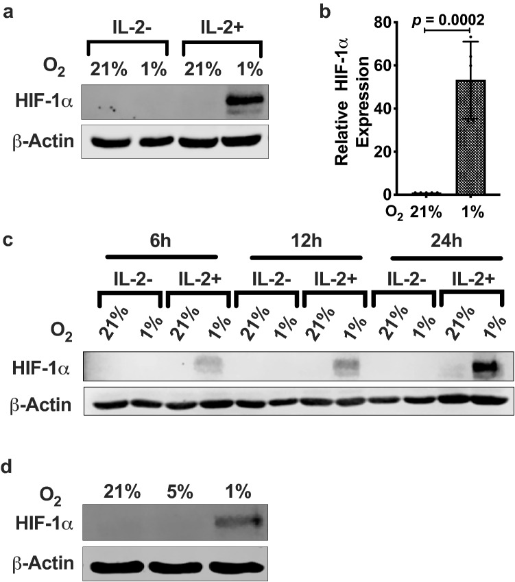
IL-2-induced HIF-1α protein expression in hypoxic NKL cells. a Immunoblot of HIF-1α expression. NKL cells were incubated in the absence or presence of 100 U/mL IL-2 for 24 h at 21% or 1% O2. Cells were lysed and immunoblotted for HIF-1α. β-Actin was used as the loading control. Blot is representative of > 4 independent trials. b Quantification of immunoblots presented as bar graphs are averages of four independent trials ± SEM. Statistical significance and p values were determined using the unpaired two-tailed Student’s t-test. p ≤ 0.05 is significant. c Time-dependent increase in HIF-1α protein levels. NKL cells were incubated in the absence or presence of 100 U/mL IL-2 for the indicated times at 21% or 1% O2. Cell lysates were immunoblotted for HIF-1α and β-Actin was used as the loading control. d Effect of physioxia (5% O2) on HIF-1α protein expression. IL-2-stimulated NKL cells exposed to 21%, 5%, or 1% O2 for 6 h were lysed and immunoblotted for HIF-1α. Data are representative of two independent trials

