Fig. 3.
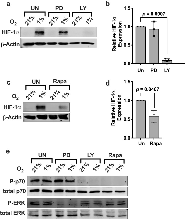
IL-2 mediates HIF-1α expression in NKL cells through the PI3K/mTOR signaling pathway. a IL-2-stimulated NKL cells were incubated for 24 h at 21% or 1% O2 in the presence or absence of 50 μM PD98059 (MAPK inhibitor) or 50 μM LY294002 (PI3K inhibitor). Lysates were immunoblotted for HIF-1α. β-Actin is the loading control. b Quantification of immunoblots normalized to β-actin is presented as fold difference of HIF-1α levels in 1% O2 relative to inhibited samples. Bar graph is representative of three independent experiments. c IL-2-stimulated NKL cells were incubated for 24 h at 21% or 1% O2 in the presence or absence of 10 nM rapamycin. Lysates were immunoblotted for HIF-1α. β-Actin is the loading control. d Quantification of immunoblots normalized to β-Actin is presented as fold difference of HIF-1α levels in 1% O2 relative to the inhibited sample. Bar graph is representative of three independent experiments. Statistical significance and p value between inhibitor treated and uninhibited were determined by unpaired two-tailed Student’s t-test. p ≤ 0.05 is significant. e IL-2-stimulated NKL cells exposed to 21% or 1% O2 were untreated or treated with 50 μM PD98059, 50 μM LY294002, or 10 nM rapamycin. Lysates were immunoblotted for the proteins as shown

