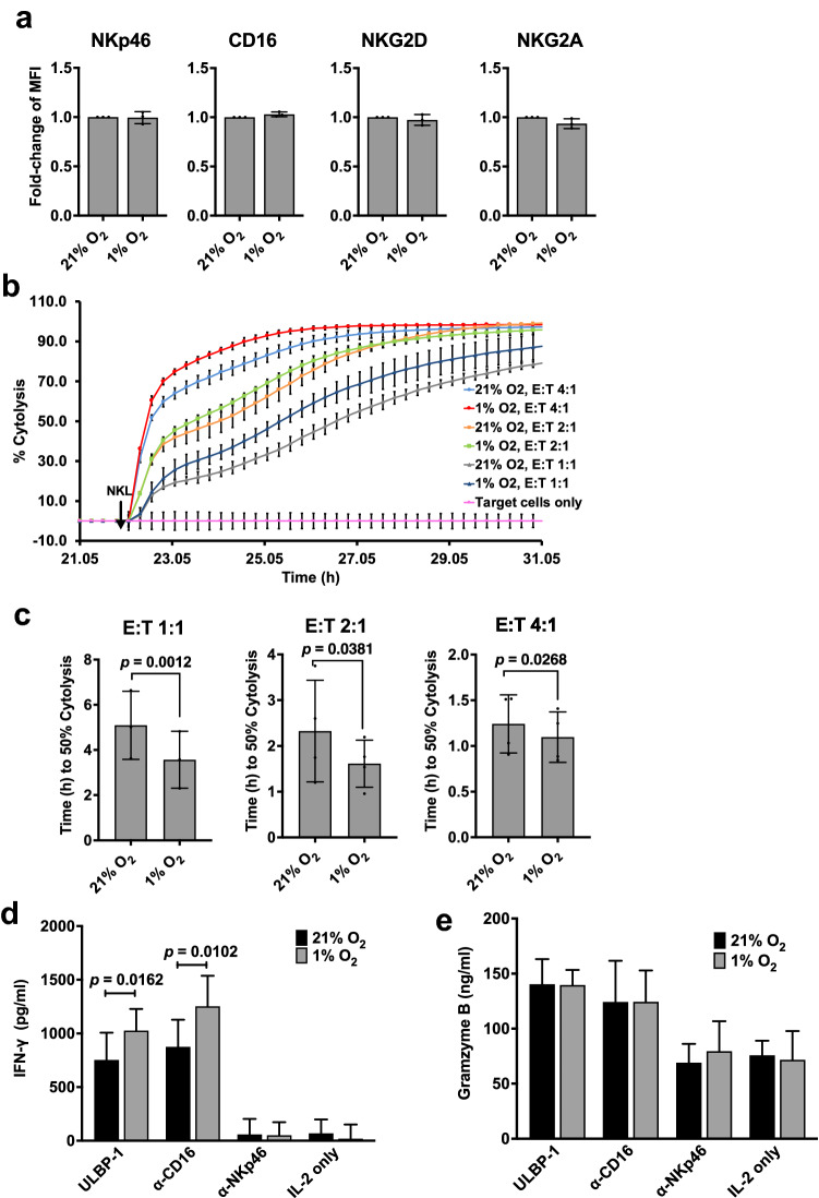
Fig. 4.
NKL cells in hypoxia exhibit increased cytolytic activity and IFN-γ secretion. a Characterization of NK cell receptors on NKL cells in hypoxia. NKL cells in the presence of IL-2 were exposed to 21% or 1% O2 for 24 h. Expression of surface receptors NKG2D, NKp46, CD16 and NKG2A was analyzed by flow cytometry as described in “Materials and methods.” Fold changes in median fluorescence intensity (MFI) ± SEM of hypoxic NKL cells against normoxic NKL cells are shown (n = 3). Statistical significance was analyzed between normoxia and hypoxia by a paired two-tailed Student’s t-test. b Real-time analysis of cytolysis exhibited by hypoxia-treated NKL cells against target tumor cells. At 24 h after seeding the target DLD-1 cells, NKL cells pre-incubated in hypoxia or normoxia in the presence of IL-2 for 24 h were added to the target cells at E/T ratios of 4:1, 2:1, and 1:1. Real-time cytolysis of target cells was monitored using the xCELLigence RTCA SP system. Electrode impedance was measured and recorded as cell index. % Cytolysis was then determined using the RTCA Software Pro. One representative of three independent experiments performed in duplicates is shown. c 50% killing time (KT 50) for the same E/T ratios in b. d NKL cells incubated at 21% or 1% O2 for 24 h in the presence of IL-2 before being transferred to plates coated with ULBP-1 or anti-NKp46 or anti-CD16 antibodies, and incubations continued under same conditions for an additional 18 h. After incubation, supernatant was collected and ELISA performed to test the concentrations of IFN-γ and granzyme B. Results are reported as the mean of four independent experiments. Statistical significance was analyzed between 21% O2 and 1% O2 using two-tailed paired Student’s t-test. p ≤ 0.05 is significant

