Fig. 5.
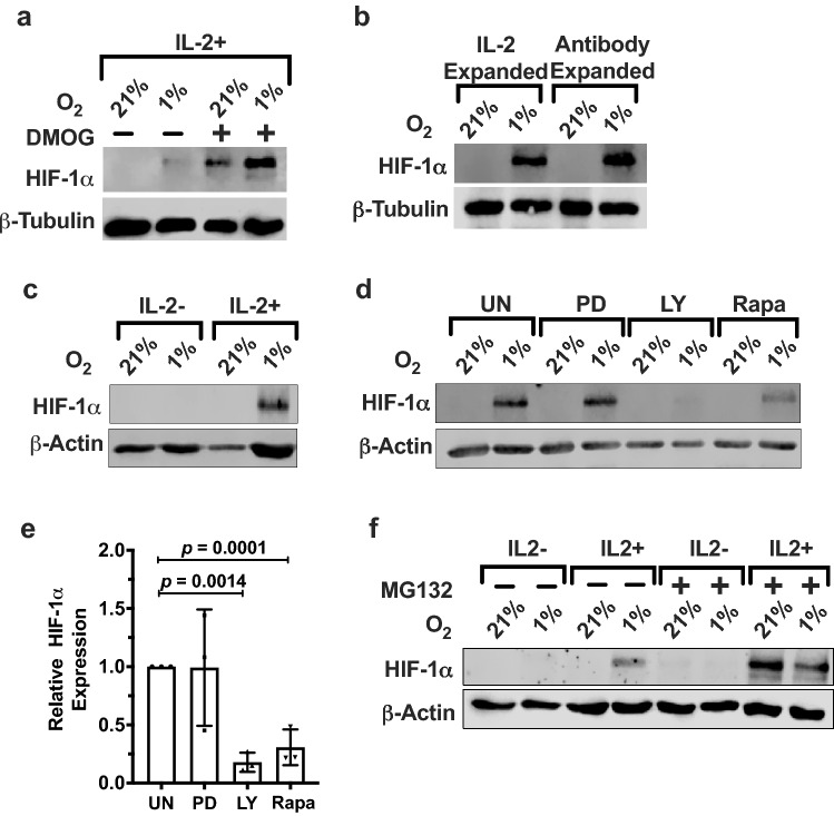
Ex vivo expanded PBMC NK cells express HIF-1α protein in the presence of IL-2. a Freshly isolated NK cells were prepared from human peripheral blood as described in “Materials and methods” and stimulated with 400 U/mL IL-2 and incubated for 72 h at 21% or 1% O2 in the presence or absence of 20 μM PHD inhibitor DMOG. HIF-1α and β-tubulin were detected by immunoblotting. Lanes lacking DMOG are representative of six different donors, and lanes with DMOG treatment are representative of three different donors. b PBMC-derived NK cells were expanded for 2 weeks using IL-2 only (IL-2 expanded) or IL-2 plus the Miltenyi MACSiBead™ loaded with anti-CD2 and anti-NKp46 abs (antibody expanded). NK cells were subsequently incubated with 400 U/mL IL-2 for 72 h at 21% or 1% O2. Whole-cell lysates were made and immunoblotted with anti-HIF-1α and β-tubulin antibodies. Blot is representative of seven different donors. c Ex vivo expanded NK cells were incubated in the absence or presence of 400 U/mL IL-2 for 72 h at 21% or 1% O2. Lysates were immunoblotted for HIF-1α. β-Actin is the loading control. d Ex vivo expanded NK cells were incubated with IL-2 for a total of 72 h at 21% or 1% O2 with the final 24 h in the presence of PD98059 (MAPK inhibitor), LY294002 (PI3K inhibitor), or rapamycin (mTOR inhibitor). Cell lysates were immunoblotted for HIF-1α. β-Actin is the loading control. Blot is representative of three independent experiments performed using three different donors. e Quantification of immunoblots in d normalized to β-actin and presented as fold difference of HIF-1α expression in 1% O2 relative to inhibited samples. Statistical significance was analyzed between inhibitor treated and control using paired two-tailed Student’s t-test. p ≤ 0.05 is significant. f Whole-cell lysates were prepared with ex vivo expanded NK cells incubated in the presence or absence of 400 U/mL of IL-2 and in the presence or absence of 10 µg/mL of MG132 for the final 24 h in a total 72-h incubation at 21% or 1% O2. HIF-1α and β-actin were detected by immunoblotting. Data are representative of two donors

