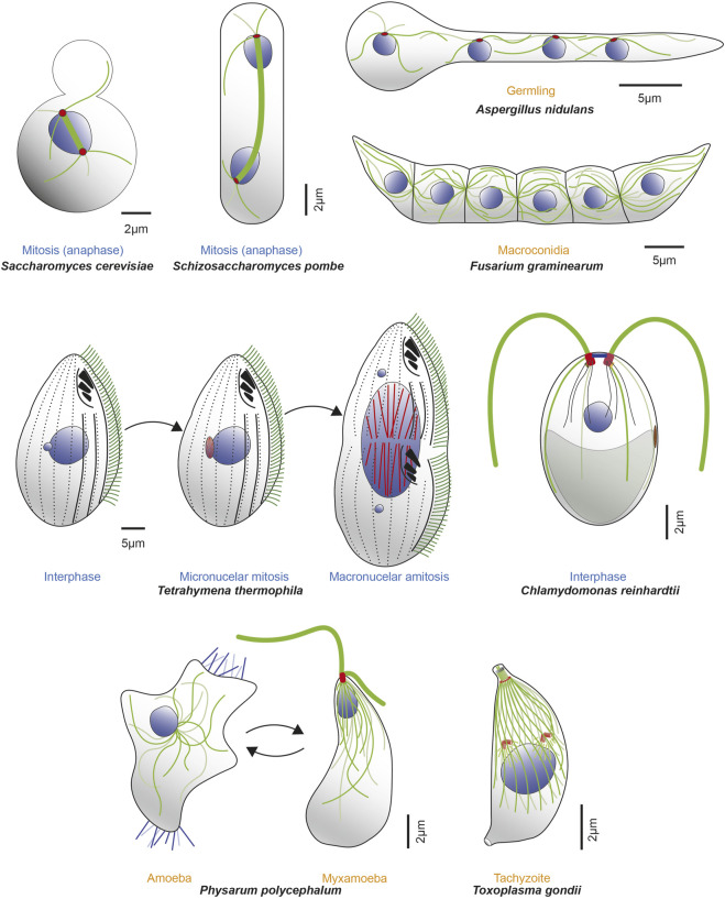
FIGURE 2.
Microorganisms express one or more α- and β-tubulin isotypes to construct various MT-organelles and to perform diverse functions with a range of complexity. Example microorganisms are represented in either different cell cycle (blue text) or life cycle (orange text) stages, along with MTs (dark and light green lines), mitotic spindle (thick green line), nuclei (blue) and corresponding MTOCs (red). Top row (from left to right, clockwise): schematics of mitotic S. cerevisiae in anaphase; mitotic S. pombe in anaphase; tetranucleate, interphase Aspergillus nidulans germling (germinated from conidia, an asexual, uninucleate spore produced during vegetative life cycle. Septa are not formed until third karyokinesis) and infectious F. graminearum multicellular macroconidia, the translucent, canoe-shaped asexual spores possessing 4–5 septa and derived from phialides, the conidium producing cells. Middle row (left): T. thermophilus undergoing cell division and demonstrating distinct localization of β-tubulin isotypes. Btu2 is enriched in somatic cilia (green lines on cell surface) and basal bodies (black dots beneath each cilium on cell surface; not all cilia are shown). Blt1 and Blt4 construct the spindle (red lines) in the mitotic division of the micronucleus (smaller blue circle) and also assemble MTs (red lines) during amitotic division of the macronucleus (large blue circle). Middle row (right): C. reinhardtii in interphase, showcasing diverse MT organelles including the two apical flagella (thick green lines projecting outward) and 4 MT rootlets (thinner green and light green lines emerging from basal bodies within the cell), which contains stable, acetylated α-tubulin. The mother (red) and daughter (dark red) basal bodies, several nucleus-basal body connectors (black lines), eyespot (brown) and chloroplast (grey object at cell posterior) are also shown. Bottom row (left): P. polycephalum depicted in respective amoeba and flagellate stages. The uninucleate amoeba (left) have unorganized MTs (green lines), randomly positioned nucleus (blue circle) with filopods (blue lines extending from the cytoplasm) that contributes to multidirectional movements. It reversibly transitions to the uninucleate, comma-shaped, flagellated, sexual spore myxamoeba (right). This phase demonstrates an anterior and posterior flagellum (thick green lines; anterior longer than posterior) that emanates from the basal body (red) as well as a flagellar cone of MTs (thinner green lines in conical arrangement, emanating from the apical basal body) that can extend to the dorsal side of the organism. The beak-shaped nucleus (blue) is positioned underneath the cone. Bottom row (right): T. gondii tachyzoite demonstrates a diverse array of complex MT-organelles. The spindle MTs (green lines) and the corset of 22 subpellicular MTs respectively nucleate from the centrioles (orange cylinders above the blue nucleus) and the Apical Polar Ring (APR) MTOC (orange circle at apical part of the cell). Also shown are the tubulin-based hollow cylindrical conoid (green cylinder above the APR), two preconoidal rings (grey circles) above it and two intraconoid MTs (green lines) within its circumference.

