
FIGURE 5.
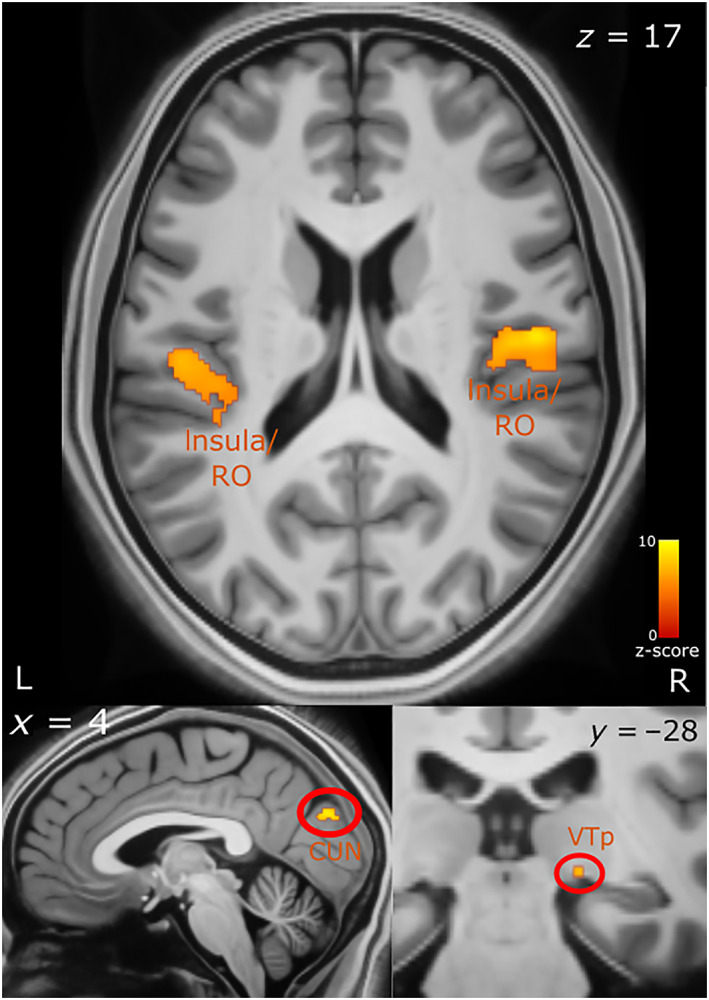
Brain activity correlated with faster response times. Top panel shows activation in the insula and Rolandic operculum. The bottom left panel shows activation in the cuneus and bottom right in the right visual thalamus. The axial (z), coronal (y) and sagittal (x) MNI coordinates are embedded in the relevant images. CUN, cuneus; L, left; R, right; RO, Rolandic operculum; VT, visual thalamus (pulvinar)