Fig. 1.

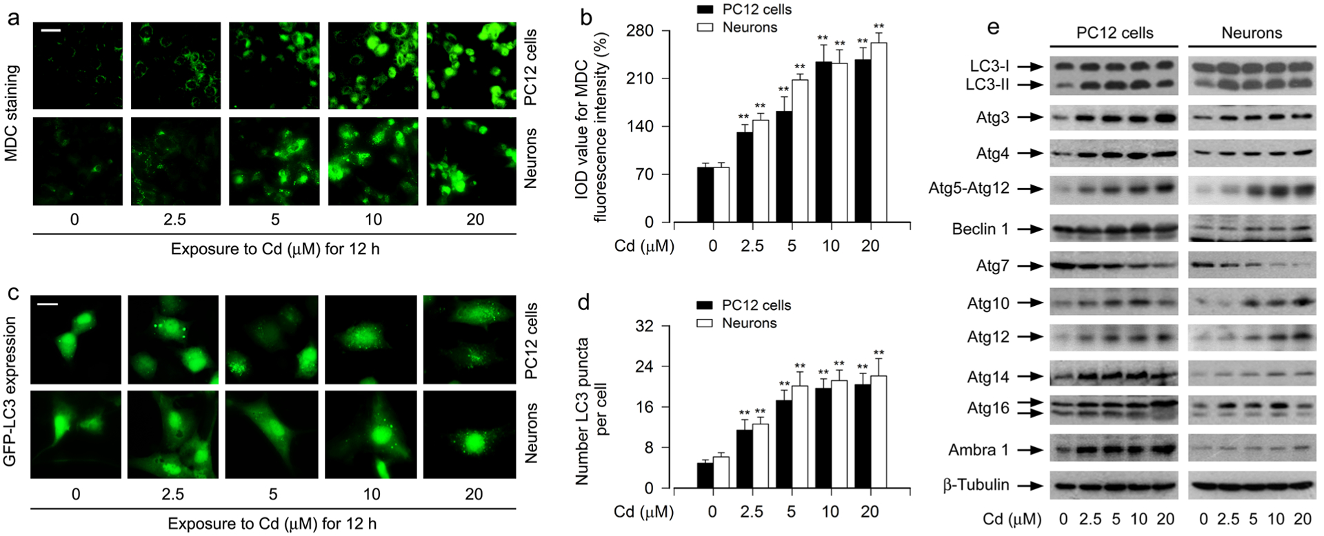
Cd induces expansion of autophagosomes with a concomitant abnormal expression of Atg proteins in neuronal cells. PC12 cells and primary neurons, or PC12 cells and primary neurons infected with Ad-GFP-LC3, respectively, were exposed to Cd (0–20 μM) for 12 h. a, b The cells were labeled using a specific autophagolysosome marker MDC staining and then the fluorescence intensity (in green) for MDC-labeled vacuoles was imaged (a) and quantified (b). Scale bar: 20 μm. c, d Shown were representative GFP-LC3 fluorescence images (in green) (c) and quantified number (d) for GFP-LC3 puncta in the cells. Scale bar: 2 μm. e Total cell lysates were subjected to Western blotting using indicated antibodies. The blots were probed for β-tubulin as a loading control. Similar results were observed in at least three independent experiments. For b and d, all data were expressed as means ± SE. n = 5, *P < 0.05, **P < 0.01, difference with control group
