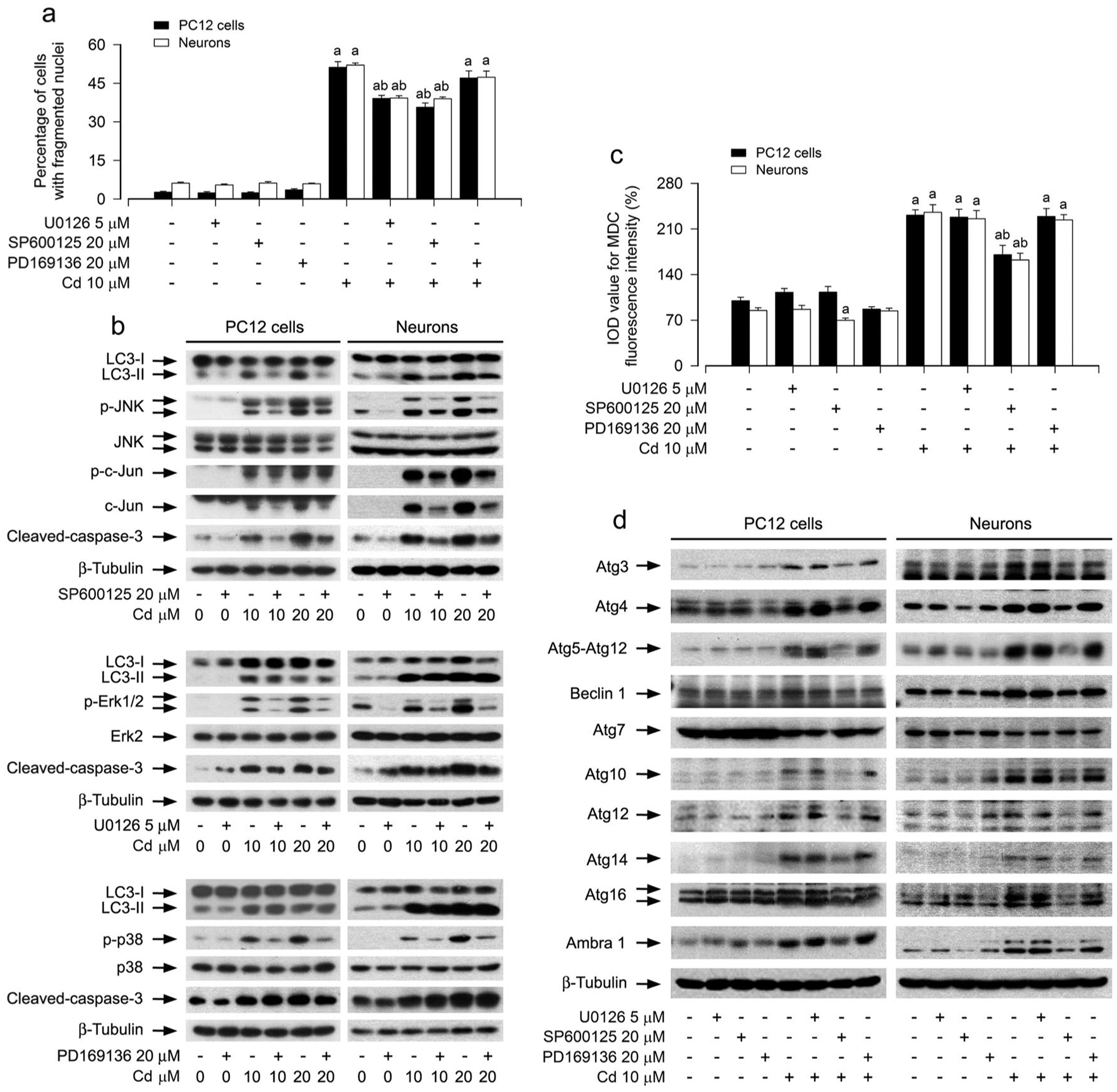
Fig. 3.

Pharmacological inhibition of JNK mitigates Cd-induced expansion of autophagosomes, abnormal expression of Atg proteins, and apoptosis in neuronal cells. PC12 cells and primary neurons were pretreated with/without U0126 (5 μM), SP600125 (20 μM) or PD169136 (20 μM) for 1 h, followed by exposure to Cd (10 μM) for 12 h (for Western blotting and MDC staining) or 24 h (for DAPI staining). a The apoptotic cells were evaluated by nuclear fragmentation and condensation using DAPI staining. b, d Total cell lysates were subjected to Western blotting using indicated antibodies. The blots were probed for β-tubulin as a loading control. Similar results were observed in at least three independent experiments. c The fluorescence intensity for MDC-labeled vacuoles in the cells was quantified. For a and c, all data were expressed as means ± SE. n = 5, aP < 0.05, difference with control group; bP < 0.05, difference with 10 μM Cd group
