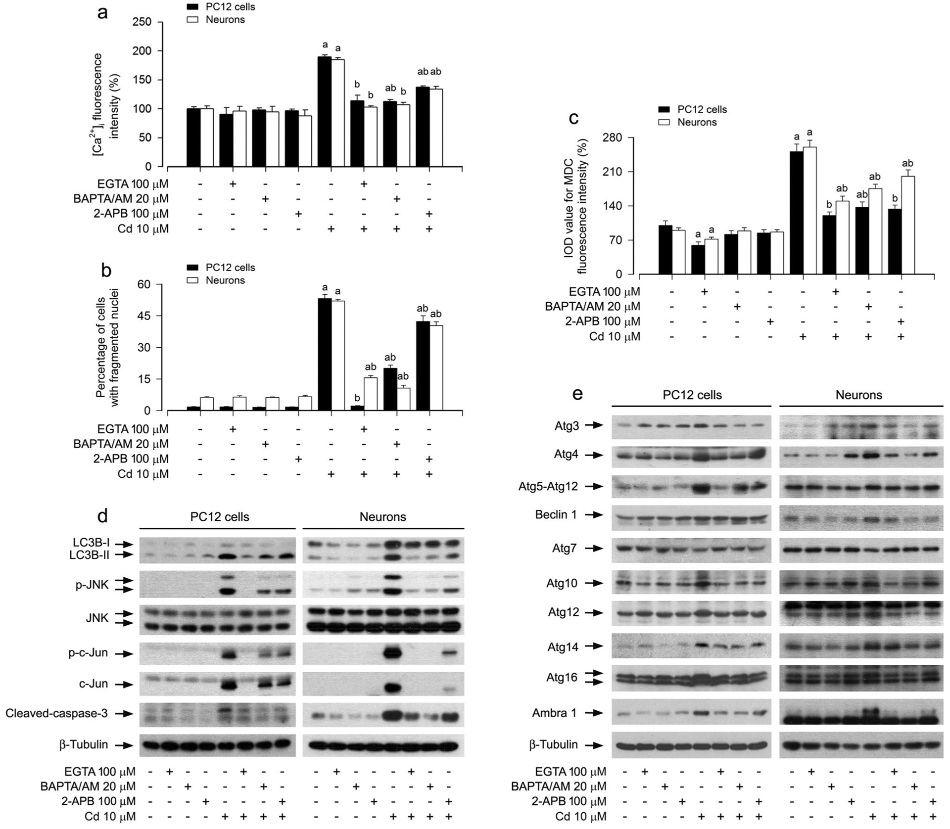
Fig. 5.

Cd activated JNK pathway contributing to expansion of autophagosomes, abnormal expression of Atg proteins, and neuronal apoptosis in a Ca2+-dependent manner. PC12 cells and primary neurons were pretreated with/without EGTA (100 μM), BAPTA/AM (20 μM) or 2-APB (100 μM) for 1 h, followed by exposure to Cd (10 μM) for 12 h (for Western blotting and MDC staining) or 24 h (for [Ca2+]i detection and DAPI staining). a [Ca2+]i manifestation was detected by a microplate reader using an intracellular Ca2+ indicator dye Fluo-3/AM. b The apoptotic cells were evaluated by nuclear fragmentation and condensation using DAPI staining. c The fluorescence intensity for MDC-labeled vacuoles in the cells was quantified. d, e Total cell lysates were subjected to Western blotting using indicated antibodies. The blots were probed for β-tubulin as a loading control. Similar results were observed in at least three independent experiments. For a, b and c, all data were expressed as means ± SE. n = 5, aP < 0.05, difference with control group; bP < 0.05, difference with 10 μM Cd group
