
Figure 6.
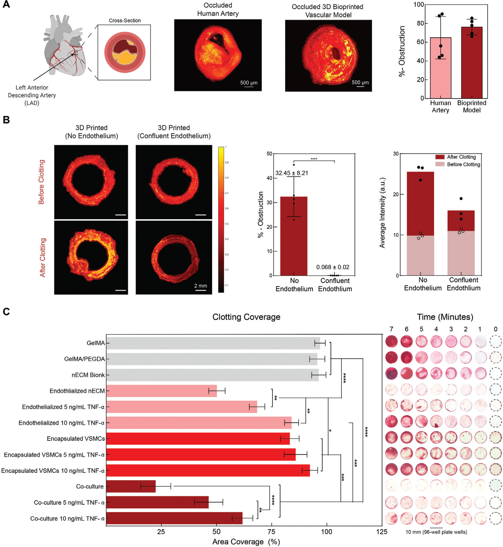
Functional validation of 3D bioprinted vascular model. A) OCT is used to determine obstruction of the lumen following blood perfusion, demonstrating the formation of diseased model. Comparing a clotted human left anterior descending artery to the occluded bioprinted vascular model (no endothelium), similar geometries are achieved and no significant difference in obstruction is observed after 12 h of clotting. B) OCT of a 3D printed (no endothelium) and 3D printed (confluent endothelium) was performed in pre- and post-stenosed models. When no endothelium was present, a significant amount of clotting is depicted, increasing the percent obstruction as compared to when a confluent endothelium is present. Comparing the average intensities between groups demonstrated a significant increase, further supporting clotting formation. C) To exemplify dose-dependent sensitivity, static clotting experiments were performed. With no lumen formation (GelMA, GelMA+PEGDA, nECM, encapsulated VSMCs, Encapsulated VSMCs with 5 ng mL−1 TNF-α, and encapsulated VSMCs with 10 ng mL−1 TNF-α), there was a significant increase in area coverage after 7 min. However upon the addition of ECs, the area coverage significantly decreases and demonstrates a dose-dependent effect on percent area coverage with TNF-α stimulation. Multiple replicates of samples were performed for rigor (n = 5) and one-way ANOVA with posthoc Turkey analysis (* = p < 0.05, ** = p < 0.05, *** = p < 0.005, and **** = p < 0.0001) was used for statistics analysis.