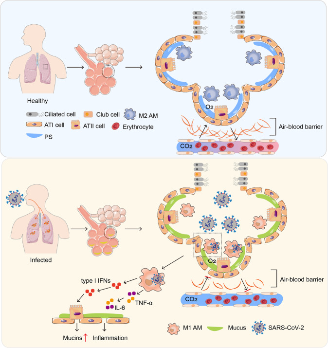
Fig. 2.
The pathological features of the lung in SARS-CoV-2-infected patients. In healthy conditions, alveolar epithelia are composed of alveolar epithelial type I (ATI) and ATII cells. ATI cells are squamous cells covering ~95% of the alveolar internal surface area; the other 5% are covered with cuboidal ATII cells, which secrete pulmonary surfactant. Alveolar macrophages (AMs) reside in airspaces, accounting for ~95% of alveolar immune cells, and safeguard against most inhaled irritants. The lung air–blood barrier acts as the site for O2 and CO2 exchange. Nevertheless, a certain amount of viruses can cause AMs polarization toward the M1 phenotype. Proinflammatory cytokines secreted by SARS-CoV-2-infected M1 AMs abrogate PS production by ATII cells, allowing the new virions released from M1 AMs to gain access to ATII cells. The type I interferons released by infected M1 AMs stimulate ATII cells to produce mucus. However, accumulation of alveolar mucus affects the blood-gas barrier, leading to impaired O2–CO2 exchange

